




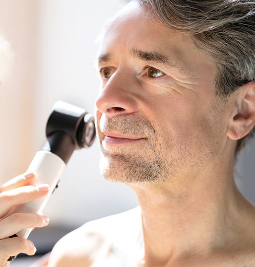

Actinic keratoses are common precancerous lesions that develop on chronically sun-exposed areas such as the scalp, face, ears, chest, and hands. They are most common in individuals with fair skin and often feel rough or scaly.
Although not all actinic keratoses progress to skin cancer, untreated lesions carry a risk of evolving into squamous cell carcinoma. Diagnosis is usually made on the basis of the physical exam alone. Treatment options are highly effective and may include liquid nitrogen, prescription topical medications, or advanced light-based treatments, depending on the extent and location of the lesions.

Cure rate when skin cancer is detected and treated in its earliest stages

Americans will develop skin cancer by age 70

UV radiation from the sun and tanning beds is the leading cause of all skin cancers

Annual skin checks are the single most effective tool for early detection and survival
Basal cell carcinoma is the most common type of skin cancer. It often appears as a pearly, pink bump, “non-healing wound”, or as a well-defined red, scaly patch on sun-exposed skin.
Risk factors include a history of sunburns, tanning bed use, fair skin, light eyes, prior burn injuries, and certain genetic conditions. Basal cell carcinomas grow slowly and rarely spread beyond the skin; however, untreated lesions can cause significant local damage. Diagnosis is confirmed with a biopsy, and treatment is individualized based on the lesion’s size, subtype, and location to achieve optimal tumor clearance and cosmetic outcomes.
Squamous cell carcinoma is the second most common form of skin cancer and is generally more aggressive than basal cell carcinoma. It may present as a scaly patch, a firm bump on a red base, or a rapidly enlarging, tender growth.
Risk factors include chronic sun exposure, fair skin, smoking, prior burn injuries, chronic inflammatory skin conditions, certain genetic disorders, and immunosuppression. A biopsy is required to confirm the diagnosis and assess the tumor’s characteristics. Treatment recommendations are tailored to the individual lesion, with careful consideration of its subtype, size, and location.
Melanoma is a form of skin cancer that arises from pigment-producing cells known as melanocytes. When identified early, melanoma is highly treatable. Delayed diagnosis, however, can lead to more advanced disease.
Melanoma may develop within an existing mole or appear on previously normal skin. It often presents as an irregularly pigmented lesion or a mole that has changed in size, shape, or color. Risk factors include fair skin, a history of sunburns, tanning bed use, numerous atypical moles, a history of melanoma, and a family history of melanoma.
Any lesion suspicious for melanoma should be promptly biopsied to confirm the diagnosis and determine tumor depth and subtype. Early detection remains the most powerful tool in achieving the best possible outcomes.
Skin cancer and premature aging are driven by cumulative ultraviolet (UV) exposure, not just severe sunburns. Sun exposure adds up over time, gradually damaging DNA, breaking down collagen, and increasing the risk of skin cancers, including melanoma.
Sun protection is one of the most effective investments you can make in your skin health. We recommend broad-spectrum sunscreen (SPF 30 or higher) every morning, with reapplication every two hours when outdoors. Sun-protective clothing, wide-brimmed hats, and sunglasses provide consistent, reliable coverage, particularly during peak UV hours (10 am to 4 pm).
Tanning beds are not a safe alternative to natural sunlight. Artificial UV radiation significantly increases the risk of melanoma and other skin cancers and accelerates premature aging. There is no such thing as a “safe tan.”
Thoughtful, consistent protection today preserves your skin’s health, strength, and appearance for decades to come.
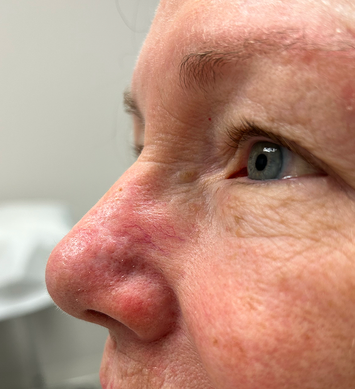
Explore before-and-after images that highlight outcomes from real patients treated in our practice. Each result reflects an individualized treatment plan developed by a board-certified dermatology team, with a focus on safety, skin health, and natural-looking improvement.
View Before & after gallery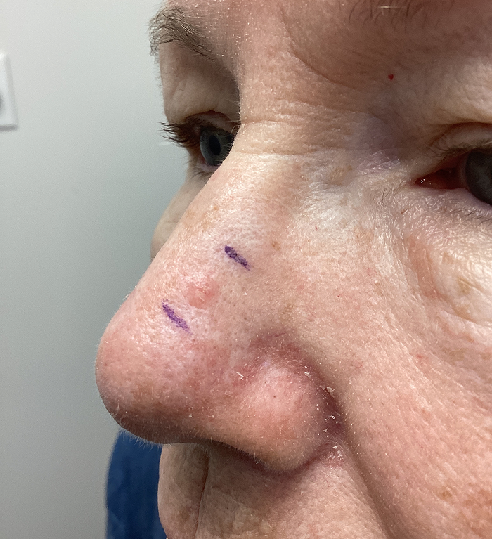





“Dr. Spring was just fabulous to deal with. She listened intently and had a great personality to boot. I’m almost 82 years old, and my initial visit with her was one of the best experiences I have EVER had with any physician. I feel privileged to have her as my doctor, and I highly recommend her!!!”
“Dr. Spring was amazing. She explained everything thoroughly and respected my concerns.”
“Dr. Spring and her staff were upbeat, attentive, and caring. They saw my anxiety prior to my procedure and took an extra moment to talk through it with me, which made me feel like they care about me as a whole person. I’ve never felt anxious prior to treatment before so this extra step really hit home.”
“Kudos to Dr. Spring. She has the Eye of an Eagle, the Heart of a Lion, yet the Hand of a Woman! Best surgeon ever!”


If you have noticed a new or changing lesion, or if it has been some time since your last skin examination, scheduling a professional evaluation is an important step in protecting your skin health. Thorough skin examinations allow for early detection of precancerous lesions and skin cancers, when treatment is most effective, and outcomes are best. To get started, schedule a consultation with Capital Skin and Laser through our online contact form. Our team will follow up to coordinate your visit and ensure your concerns receive the attention and care they deserve.
"*" indicates required fields
